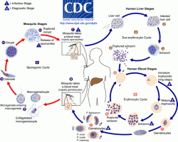
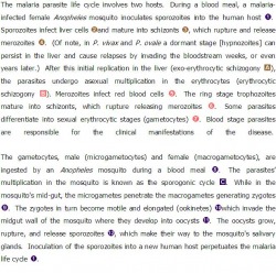
Malaria Life Cycle
Plasmodium vivax
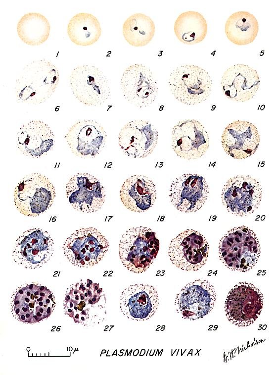
Morphology
Relative age of infected RBCs Only those that are young & immature
Appearance of infected RBCs Enlarged, distorted
Relative age of infected RBCs Only those that are young & immature
Appearance of infected RBCs Enlarged, distorted

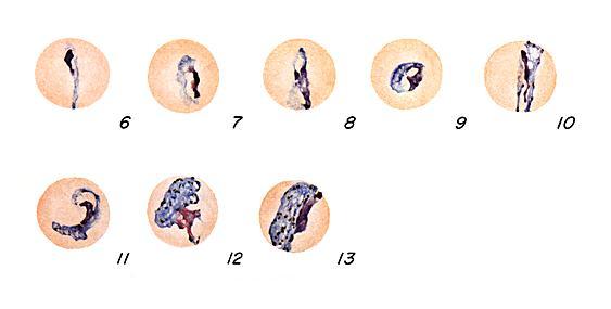
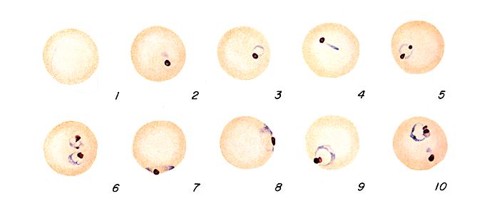
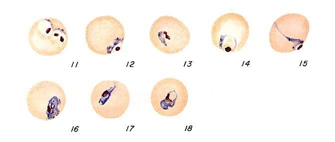
Ring Form
Ring Form:
- Delicate cytoplasmic ring measuring 1/3 RBC diameter
- Single chromatin dot
- Ring surrounds a vacuole
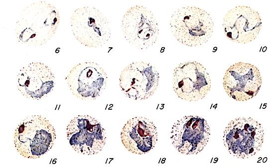
Trophozoite
Trophozoite:
- Irregular ameboid appearance
- Ring remnants common
- Brown pigment
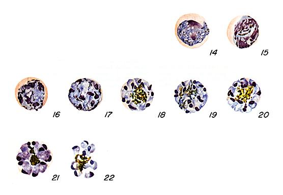
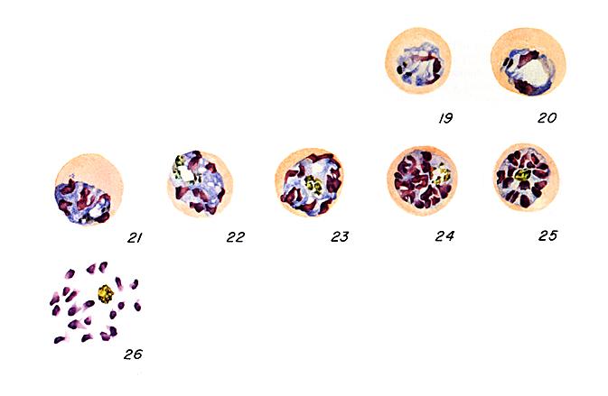
Schizont
Schizont:
Immature schizont
Immature schizont
- Multiple chromatin bodies
- Brown pigment
- 12 to 24 merozoites occupying majority of the red cells
- Merozoites surrounded by cytoplasmic material
- Brown pigment may be present
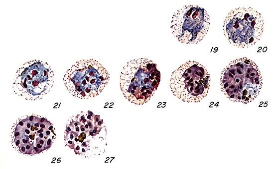
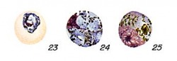
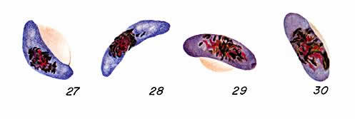
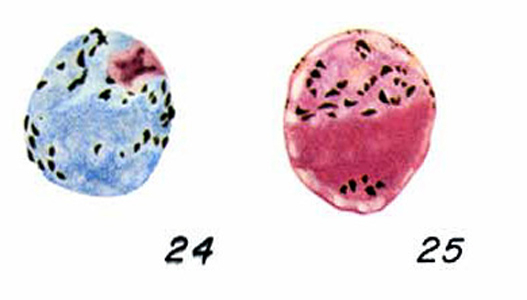
Gametocyte
Gametocyte:
Microgametocyte
Microgametocyte
- Large pink to purple chromatin mass surrounded by colourless to pale halo
- Brown pigment common
- Round to oval cytoplasm
- Eccentric chromatin mass
- Delicate light-brown pigment present throughout cell
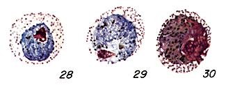
Plasmodium ovale
Morphology
Relative age of infected RBCs Only those that are young & immature
Appearance of infected RBCs Oval & enlarged, distorted with ragged
cell walls
Relative age of infected RBCs Only those that are young & immature
Appearance of infected RBCs Oval & enlarged, distorted with ragged
cell walls
Ring form:
- Resembles that of P. vivax
- Ring larger in size than P. vivax
- Ring often tick & somewhat ameboid in appearance
Ring Form
Trophozoite:
- Ring appearance usually maintained until late in development
- Ameboid tendencies not as evident as P. vivax
Trophozoite
Schizont:
Immature schizont
Immature schizont
- Progressive dividing chromatin surrounded by cytoplasmic material, often maintains circular shape early in development
- Parasites occupy three quarters of RBCs
- 8 to 12 merozoites maybe present
- Rosettes of an average of 8 merozoites

Schizont
Gametocyte:
- Similar to P. vivax, only smaller in size
Gametocyte
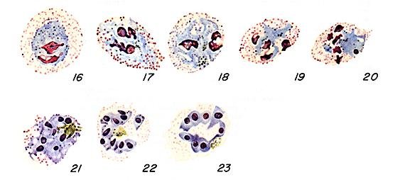
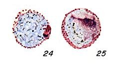
Plasmodium malariae
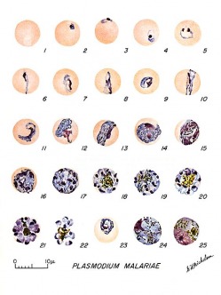
Morphology
Relative age of infected RBCs Only mature cells
Appearance of the infected RBCs Normal size, no distortion
Relative age of infected RBCs Only mature cells
Appearance of the infected RBCs Normal size, no distortion
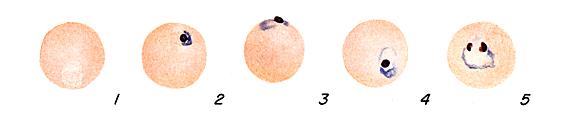
Ring Form
Ring form:
- Smaller in size than P. vivax
- Occupies 1/6 of the RBS
- Heavy chromatin dot
- Vacuole may appear “filled in”
- Pigment forms early
Trophozoite
Trophozoite:
- Non-ameboid solid cytoplasm that may assume a roundish, oval, band, or bar shape
- Cytoplasm contains coarse dark brown pigment that may mask chromatin material
- Vacuoles are absent in mature stages
Schizont
Schizont:
Immature schizont
Immature schizont
- Similar to that of P. vivax, only smaller and may contain large & dark peripheral or central granules
- 6 to 12 merozoites arranged in rosettes or irregular clusters
- Central arrangement of brown-green pigment may be visible
Gametocyte:
- Similar to P. vivax, only smaller in size & pigment is usually darker & more coarse
- Older forms assume an oval shape
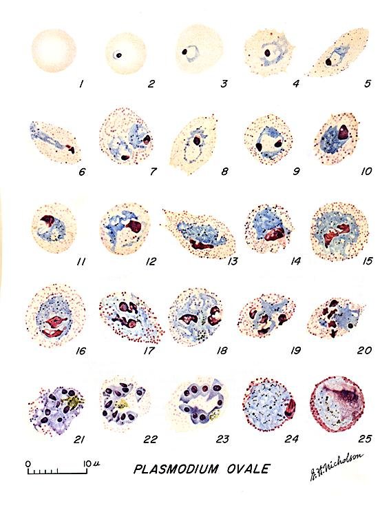
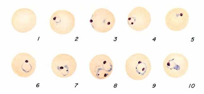
Plasmodium falciparum
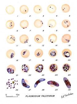
Morphology
Relative age of infected RBCs May infect cells of all ages
Appearance of infected RBCs Normal size, no distortion
Relative age of infected RBCs May infect cells of all ages
Appearance of infected RBCs Normal size, no distortion
Ring form:
- Circle configuration (one chromatin dot) or headphone configuration (two chromatin dots)
- Scanty cytoplasm & small vacuole
- Multiple rings common
- Accolé forms common
Ring Form
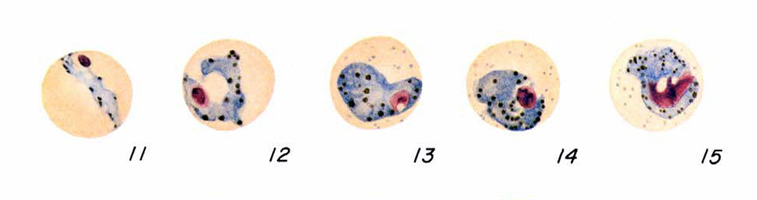
Trophozoite:
- “Heavy rings” common with fine pigment granules
- Mature forms only seen in severe infections
Trophozoite
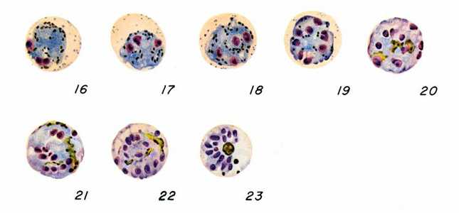
Schizont:
Immature schizont
Immature schizont
- Multiple chromatin bodies surrounded by cytoplasm
- Only detected in severe infection
- 8 to 36 merozoites (average 24) in clusters arrangement
- only detected in severe infections
Schizont
Gametocyte:
Microgametocyte
Microgametocyte
- Sausage or crescent shape
- Dispersed central chromatin with nearby black pigment visible
- Sausage or crescent shape
- Compact chromatin
- Black pigment visible
Gametocyte
Plasmodium knowlesi
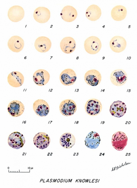
Morphology
Appearance of infected RBCs Normal size, no distortion
The appearance of P.knowlesi resembles those of P.falciparum
Appearance of infected RBCs Normal size, no distortion
The appearance of P.knowlesi resembles those of P.falciparum
Ring form:
- Delicate cytoplasm
- 1 to 2 prominent chromatin dots
- Occasional appliqué (accolé) forms
Trophozoite:
- Compact cytoplasm
- Large chromatin
- Occasional band forms
- Coarse, dark-brown pigment
Schizont:
- Mature = up to 16 merozoites with large nuclei
- Clustered around mass of coarse, dark-brown pigment
- Occasional rosettes
- Mature merozoites appear segmented
Gametocyte:
- Round to oval
- Compact, may almost fill RBC
- Chromatin compact
- Eccentric (macrogametocyte)
- More diffuse (microgametocyte)
- Scattered brown pigment
